Reproductive Genetics Clinic

Prenatal screening
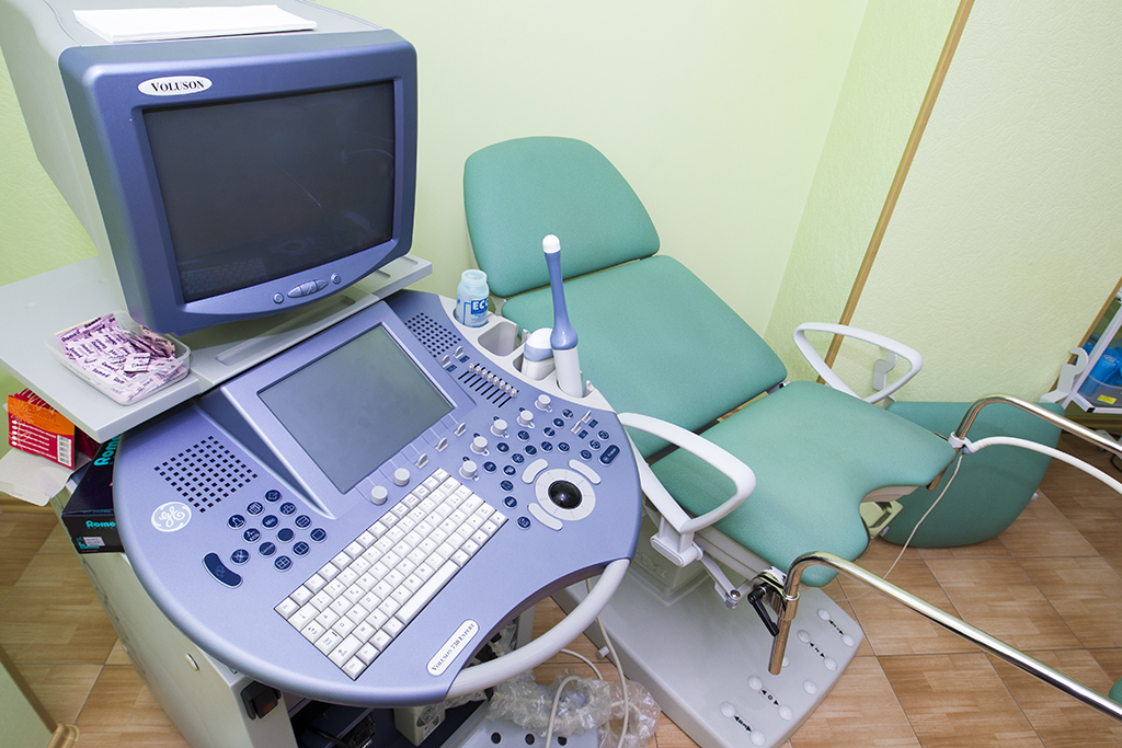
Prenatal diagnostics allows to assess the state of health of an unborn child and, in case of deviations, pay attention to them in time. The “Victoria” clinic offers a prenatal examination in one day thanks to the successful combination of highly qualified doctors of prenatal medicine, specialists in cytogenetic and laboratory diagnostics.
Prenatal Screening and Diagnosis
Prenatal testing is a comprehensive examination of a pregnant woman, which allows determining the degree of risk of developing congenital and hereditary defects in the fetus. The “Victoria” Clinic specialists recommend screening for all pregnant women, regardless of age and the presence of hereditary diseases.
Our specialists diagnose the condition of the fetus at an early stage of development in order to provide the family with timely information about the course of pregnancy, possible complications, and signs of chromosomal and other congenital and hereditary anomalies.
Screening examinations only help to form risk groups for further research, and are not a diagnosis.
How is the examination of the fetus and pregnant woman carried out?
I trimester
• a pregnant woman undergoes an ultrasound examination during the period of 11-13 weeks of pregnancy. This is an expert fetal ultrasound examination performed by a prenatal specialist to identify markers for fetal chromosomal abnormalities or pregnancy complications. If the gestational age is informative for calculating the risk, the pregnant woman is offered to donate blood to determine the concentration of biochemical markers (PAPP-A and β-hCG);
• the individual risk of chromosomal syndromes is calculated by a special program based on fetal ultrasound data, indicators of biochemical markers (PAPP-A and β-hCG) and other characteristics of a pregnant woman;
• according to the results of these calculations, the pregnant woman is referred to the group of low (93%), medium (5%) or high (2%) risk, which is indicated in the conclusion of prenatal testing;
• further, the geneticist consults and explains to the pregnant woman the result obtained. The same doctor who performed the sonography communicates with the pregnant woman in the future, explains the indicators and gives recommendations for further research.
II trimester
• our clinic offers a unique opportunity to provide ULTRASOUND expert examination of the fetus paired with the variance analysis of biochemical markers in the second trimester (AFP, HCG and free estriol) and calculate the risk with correction of ultrasound markers of fetal pathology. This test is undertaken between 16-18 weeks (it is permitted to be done between 15-21 weeks).
Important!
Informativity of examination of the fetus between 11-13 weeks is significantly higher than in the second trimester, so we don’t recommend you to re-calculate the risk, having a high-risk pregnancy in the first trimester, instead apply for invasive prenatal testing.
Non-invasive genetic testing (by the blood of the pregnant)
Our experts are using most innovative and advanced technologies proven by the leading scientists in their work. One of this methods is a non-invasive test (by the pregnant’s venous blood) to identify the RH factor of the fetus, trisomy of chromosomes 21, 18, 13 and sex of the fetus (NIPT).
The test result is displayed in the form of risk and in most cases is almost well defined: the risk of trisomy of>99% (need to confirm fetal chromosomal abnormalities using invasive procedures) or <1% (99% chance that the fetus does not have this trisomy). Results of non-invasive tests should be interpreted by the geneticist.
Who is recommended non-invasive genetic testing?
- Pregnant with Rh-negative blood type in order to take measures to prevent rhesus-conflict timely, if the fetus was defined with positive RH factor.
- If there is a high risk of the disease, which is inherited by the gender.
- While detecting a high risk of Down syndrome (trisomy 21), Edwards (trisomy 18) or Patau (trisomy 13) for women who are having difficulties to make a decision to undertake invasive prenatal diagnosis.
When is non-invasive genetic testing done?
- The gene of the Rhesus factor (RHD) can be identified in the blood of a pregnant starting about 12 week of pregnancy with a high probability of (99%).
- The NIPT test is made from 10 week of pregnancy.
Invasive Prenatal Diagnosis
Invasive interventions are performed only if there are indications that are regulated by the current orders of the Ministry of Health of Ukraine, starting from 11 weeks of pregnancy.
The “Victoria” Clinic specialists have many years of experience in invasive interventions and carry out genetic analysis of the material in accordance with the recommendations of the European Cytogenetic Association.
Invasive prenatal diagnostics includes
chorionic (placenta) biopsy, amniocentesis and cordocentesis.
• Chorionic biopsy examination is carried out from 11 weeks to 14 weeks; placenta biopsy - from 16 weeks up to 20 weeks, in connection with maturation of the placenta. The results take from 3 to 14 days.
• Examination of amniotic fluid cells (amniocentesis) is performed from 16 to 22 weeks of pregnancy. Its advantages are the possibility of obtaining high quality chromosome preparations and the parallel conduct of biochemical or immunological studies. The results take from 2 to 3 weeks.
• The study of cord blood lymphocytes (cordocentesis) is carried out from the 19th week of pregnancy (carried out only in the group of pregnant women at high risk of fetal chromosomal abnormalities).
An invasive prenatal diagnostics is prescribed if as follows.
• When there is a high level of fetal chromosomal abnormality after combined screening.
• If congenital malformations or ultrasound markers of fetal chromosomal pathology are found during the pregnancy.
• If the age of the pregnant woman is 35 years or more (recommended by the WHO, although combined screening allows such women to determine the individual risk of fetal chromosomal abnormalities).
• If there were chromosomal abnormalities of a fetus or a newborn in previous pregnancies.
• If the future parents have chromosomal rearrangements.
Invasive method can be employed if following tests are done:
- general blood and urine tests;
- blood group and Rh factor (if negative, anti-Rh antibodies should be determined)
- coagulogram;
- determination of antibodies to HIV, hepatitis B and C;
- blood glucose;
- blood test for RW;
- pregnancy ultrasound;
- microscopy of the urogenital smear.
The risk of complications during invasive diagnosis
Any surgical interference is always associated with the risk of complications. Risks with invasive procedures are uncommon, and proven that the complication rate depends on the proper determination of gestational age and experience of the doctor.
The longstanding experience of our specialists allows minimizing the risks. The frequency of complications after invasive diagnostics in the “Victoria” clinic is not greater than 0.05% when the efficiency is 99.6% and a detection rate of chromosomal abnormalities is 13-15%.